Researchers Develop a Graphene Based Sensor to Visualize Electric Fields
As silicon-based fabrication technology is reaching its operational limits, there is increasing research attention towards developing circuits with 2D materials. The 2D materials are crystals consisting of a single layer of atoms, with their thickness ranging from a single to a few-atomic layers thick.
As silicon-based fabrication technology is reaching its operational limits, there is increasing research attention towards developing circuits with 2D materials. The 2D materials are crystals consisting of a single layer of atoms, with their thickness ranging from a single to a few-atomic layers thick. In conventional materials, effects such as quantum tunneling become prominent as the feature lengths decrease. Therefore, 2D materials such as graphene are considered as a potential candidate for the next generation of electronic devices.
2D materials exhibit exceptional chemical and physical properties including layered structure, high-surface-area, layer-dependent, optical bandgap, and changes in chemical compositions. Due to these properties, 2D materials have improved properties and detection limits, which are very critical when sensitivity is involved.
Researchers at the University of California at Berkeley and Stanford University have developed a graphene “camera” to detect a beating heart.
Currently, for recording heartbeats, chemical dyes, and electrodes are used, which can record voltages at a single measurement point, with a sheet capable of measuring the voltage over an entire surface at once. But with their camera made from a sheet of graphene, the researchers can image all cells simultaneously, and they don’t have just a point measurement, and therefore, they don’t have to scan. With their developed sensor, they can image the entire network of cells at the same time.
“The ease with which you can image an entire region of a sample could be especially useful in the study of neural networks that have all sorts of cell types involved,” says fellow first author Allister McGuire, PhD. “If you have a fluorescently labeled cell system, you might only be targeting a certain type of neuron.”
He further added,
“Our system would allow you to capture electrical activity in all neurons and their support cells with very high integrity, which could really impact the way that people do these network level studies.”
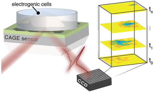
The developed critically coupled waveguide-amplified graphene electric field (CAGE) sensor uses the field-sensitive optical transitions in graphene to convert electric potentials into the optical regime. To get real-time visualization of electric fields over an area, the sample needs to be placed on the top of a graphene sheet, which is placed above a waveguide. The laser is incident on the waveguide, through a prism, and reflects off the graphene. This makes the electric field visible in real-time.
The researchers tested their sensor on a chicken heart. They used the sensor to detect native electrical activity from cardiac action potentials with a tens-of-microns resolution, simultaneously map the propagation of these potentials at tissue-scale, and monitor their modification by pharmacological agents.
“One of the things that is amazing to me about this project is that electric fields mediate chemical interactions, mediate biophysical interactions — they mediate all sorts of processes in the natural world — but we never measure them,” says Balch. “We measure current, and we measure voltage,” Balch said. “The ability to actually image electric fields gives you a look at a modality that you previously had little insight into.”
The work is described in the journal ACS Nano Letters.


